Special stainings
Special Histochemical Staining
Once we have thin, high quality sections mounted on glass slides, we can perform a variety of Special Histochemical staining to highlight specific elements of interest. Our team is highly experienced in the optimization and application of special stains for selective staining of unique tissue elements of interest.
Below is a sampling of the types of staining methods we perform:
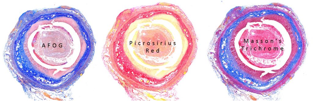
- Masson Trichrome (MT) staining: This staining method is mainly used to distinguish collagen from muscle tissue, applicable and recommended in fibrosis models, wound healing studies, infarct models and others.
- Periodic acid–Schiff (PAS) staining: This staining method is used for staining of polysaccharides such as glycogen, and mucosubstances such as glycoproteins, glycolipids and mucins in tissues.
- Toluidine blue staining: This method is used for staining of mast cells that are found in the connective tissue and their cytoplasm contains granules composed of heparin and histamine. Following Toluidine staining, mast cells are stained red-purple and the background is stained blue.
- Oil Red O: Oil Red O stain is based on the use of a lysochrome (lipo-soluble dye) and an azo dye used for the visualization of neutral triglycerides and lipids on frozen tissue sections.
- And many more!
For custom staining methods, contact our team to discuss and we will accommodate your research needs.
Special Staining list available here